Biomedical ultrasound applications
Ultrasound has important biomedical applications in both, therapy and imaging. I'm developing several biomedical ultrasound applications including focused transcranial ultrasound devices based on metamaterials and protocols for the treatment of neurological disorders; hybrid ultrasound imaging techniques, combining ultrasound with additional physical mechanisms to go a step beyond standard US imaging techniques; and ultrasonic technology to the field of odontology.
Lines:
Transcranial propagation of therapeutic ultrasound

Focused ultrasound has demonstrated spectacular capabilities for the treatments of neurological disorders as well to open new paths to deliver drugs in the central nervous system. However, focusing through the skull supposes a challenge due to the high acoustic impedance contrast, refraction index and absorption that present the skull bones. I'm interested in developing new tools to focalize and control acoustic beams into de central nervous system. These research lines include:
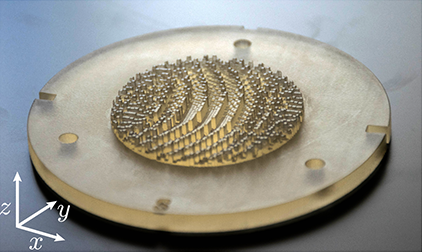
Holograms and metasurfaces for transcranial focusing: In this research line we develop passive focusing methods based on acoustic holograms to produce focused beams inside the central nervous system (CNS). In particular, we are able to produce beams whose spatial distribution fits a target CNS structure of arbitrary shape. In this way, using 3D-printed holographic lenses at UMIL we are developing low-cost focusing systems that will help to disseminate incoming therapeutic ultrasound techniques as neuromodulation or blood-brain barrier opening to treat neurological disorders.
Transcranial ultrasound for blood-brain barrier opening: The blood-brain barrier (BBB) restricts the diffusion of microscopic objects, e.g., protecting the brain from infections. However, it also prevents the passage of most therapeutic drugs. BBB disruption can be achieved by using transcranial focused ultrasound and microbubble injection in an effective, non-invasive, transient, localized and safe manner. Thus, the BBB opening enables drug delivering to specific areas of the brain, which is mandatory in research of treatments for neurological disorders as Alzheimer’s or Parkinson’s diseases. At the UMIL we develop simulations and experiments to define a protocol for the transcranial targeting of the hippocampus of an adult human by using a single-element focused transducer. In this line I'm working at the Ultrasound Medical and Industrial Laboratory (UMIL)Universitat Politècnica de València (Spain) and Columbia University (USA) to study the acoustic aberrations produced in transcranial propagation and methods to correct them. We develop simulations and experiments to define a protocol for the transcranial targeting at human hippocampus by using a single-element focused transducers.
In the right animation you can see an example of the transcranial beam propagation generated by a mono-element 500 kHz transducer, obtained by numerical simulation by PhD student Sergio Jiménez-Gambín who is working in this topic. The simulations, based on the k-space method, allow to evaluate the beam aberrations and predict precisely the central nervous system area to be treated.
Sergio Jiménez-Gambín and Noé Jiménez and José María Benlloch and Francisco Camarena,
Physical Review Applied, 12 (1), pp 014016, (2019)
Marcelino Ferri and José María Bravo and Javier Redondo and Sergio Jiménez-Gambín and Noé Jiménez and Francisco Camarena and Juan Vicente Sánchez-Pérez,
Polymers, 11 (9), pp 1521, (2019)
N. Jiménez, F. Marquet, F. Camarena, E. E. Konofagou, J. Redondo, B. Roig, R. Picó,
Revista de Acustica, 43 (1), pp 5-11, (2012)
P. C. Iglesias, N. Jiménez, E. Konofagou, F. Camarena, J. Redondo
Physics Procedia., 63, pp 103-107, (2014)
Hybrid ultrasonic imaging techniques

At the UMIL of the I3M we develop hybrid ultrasound imaging techniques, combining ultrasound with additional physical mechanisms to go a step beyond standard US imaging techniques. These include:
Magneto-Motive ultrasound imaging detects the presence of superparamagnetic nanoparticles through their mechanical responses to an external transient magnetic excitation. Due to their weak diamagnetic properties, normal tissue constituents do not respond to the magnetic field. However, when tissue is labeled with magnetic nanoparticles, it tends to move towards areas of lower magnetic potential. The magnetically-induced displacement within nanoparticles and the tissue associated with them is detectable by ultrasound imaging. At UMIL we are already developing a first prototype for magnetic nanoparticle detection.
Photoacoustic imaging techniques: When an ultrashort and intense laser illuminates light absorbing materials, a part of the absorbed energy produces locally ultrasonic pulses due to thermoelastic effect. These signals are detected and can be used to reconstruct an image that take into account the absorbance of the tissue. In this way, as the absorbance spectrum varies between tissues, it enables the ultrasonic imaging at molecular level.
Ultrasonic elastography: Ultrasonic elastography is a well-established diagnostic tool for pathologies such as liver fibrosis. At the UMIL we are developing novel elastographic techniques using acoustic radiation force and hybrid techniques.
Ultrasonic technology applied to odontology

In collaboration with the Valencian Institute of Dental Research (IVIO), within the framework of IVIO-UPVChair, we develop new ultrasonic techniques for monitoring, diagnosis and treatment in the field of dentistry. These research lines include:
Monitoring guided-bone regeneration during implantation: In particular, we develop ultrasonic characterization methods for guided bone regeneration during implantation in odontology. We aim to characterize the complete regeneration process using ultrasound to guide the treatment, optimize implantation and avoid or detect complications. The use of ultrasound is desirable as it is low cost and non-ionizing radiation, thus, it can be employed to monitor long healing treatments without the risks associated to X-Ray Computed Tomography imaging techniques.
Ultrasonic characterization of teeth and synthetic bone-grafts: Moreover, ongoing research is developed at UMIL to quantitatively characterize the mechanical properties of teeth, e.g., demineralization, and synthetic bone-graft materials using ultrasonic techniques.
Josep Rodríguez-Sendra and Noé Jiménez and Rubén Picó and Joan Faus and Francisco Camarena,
IEEE Transactions on Ultrasonics Ferroelectrics and Frequency Control, 66 (10), pp 1658-1666, (2019)