Therapeutic ultrasound and acoustic holograms
Focused ultrasound has demonstrated spectacular capabilities for the treatments of neurological disorders as well to open new paths to deliver drugs in the central nervous system. However, focusing through the skull supposes a challenge due to the high acoustic impedance contrast, refraction index and absorption that present the skull bones. I'm interested in developing new tools to focalize and control acoustic beams into de central nervous system. These research lines include:
Lines:
Holograms for transcranial focusing
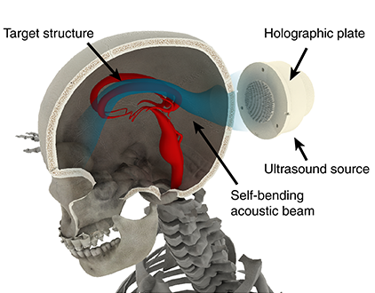
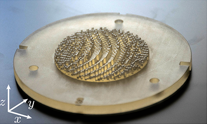
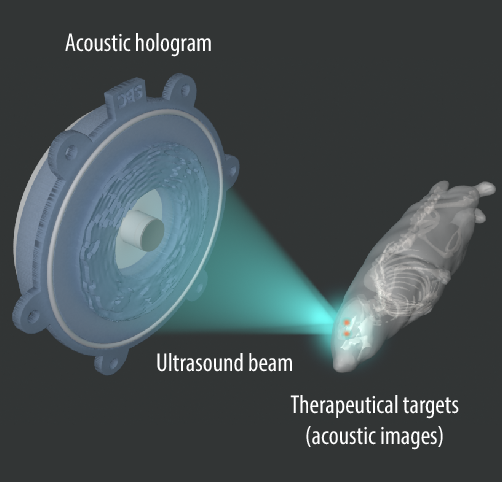
In this research line we develop passive focusing methods based on acoustic holograms to produce focused beams inside the central nervous system. In particular, we are able to produce beams whose spatial distribution fits a target structure of arbitrary shape, such as deep-brain nuclei. In this way, using 3D-printed holographic lenses we are developing low-cost focusing systems that will help to disseminate incoming therapeutic ultrasound techniques as neuromodulation or blood-brain barrier opening to treat neurological disorders.
Currently we have validated this technique in vivo, opening the blood-brain barrier in small animals in symetrical regions in separate hemispheres. In addition, holograms are able to encode the multiple reflections inside the cranial walls, increasing the angular spectrum of the focused beams. This result in sharper focal spots, that can be as narrow as the diffraction limit.
S Jiménez-Gambín, N Jiménez, AN Pouliopoulos, JM Benlloch, EE Konofagou, F Camarena,
IEEE Transactions on Biomedical Engineering, 69 (4), pp. 1359-1368, (2022)
D Andrés, N Jiménez, JM Benlloch, F Camarena,
Ultrasound in Medicine and Biology, 48 (5), pp. 872-886, (2022)
Sergio Jiménez-Gambín and Noé Jiménez and José María Benlloch and Francisco Camarena,
Physical Review Applied, 12 (1), pp 014016, (2019)
Marcelino Ferri and José María Bravo and Javier Redondo and Sergio Jiménez-Gambín and Noé Jiménez and Francisco Camarena and Juan Vicente Sánchez-Pérez,
Polymers, 11 (9), pp 1521, (2019)
Vortices in the brain

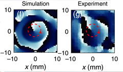
Acoustic vortex beams have great potential for contactless particle manipulation and torque-based biomedical applications. However, when focusing acoustic waves through aberration layers such as the human skull at ultrasonic frequencies results in strong phase aberrations which prevent the generation of sharp acoustic images. In the case of a wavefront containing phase dislocations, skull aberrations inhibit the focusing of acoustic vortex beams inside the cranial cavity.
We demonstrate that phase-conjugated acoustic holograms can encode time-reversed fields simultaneously allowing the compensation of the aberrations of the skull and the generation of a focused vortex inside an ex-vivo human skull. The method is applied for single-element geometrically focused sources and results in a very simple and compact ultrasonic system. The technique was tested using an exvivo skull inside a water tank. Measurements show that a vortex was correctly synthesized using an acoustic hologram. This technique can be used to design low-cost particle trapping applications, clot manipulation, torque exertion in the brain and acoustic-radiation-force based biomedical applications.
S Jiménez, N Jiménez, F Camarena,
Physical Review Applied, 14 (5), pp. 054070, (2020)
Holographic thermal patterns for hyperthermia

Holograms can shape wavefronts to produce arbitrary acoustic images. Compared to light, acoustic waves can penetrate much deeper into biological media. Therefore, acoustic holograms are bursting onto current biomedical applications due to their capability to shape and focus ultrasound fields inside living tissues. Acoustic waves transport energy, and when propagating into biological media, a fraction is irreversibly transformed into heat by viscoelastic and relaxation processes. In ultrasound imaging applications, the temperature rise is negligible because the total energy of the wave is moderate.27 However, at high ultrasound intensities, heat deposition can be elevated and fast enough to create cell necrosis and tissue ablation. Combined with sharp focusing, high intensity focused ultrasound is currently used in clinics for therapeutical purposes.

Recently, attention has been paid to intermediate regimes, when ultrasound is used to create a local and mild hyperthermia, which is defined as an increase in the tissue temperature between 39 and 45C, typically with long exposure times.30–32 HIFU-mediated hyperthermia is emerging as a highly promising therapeutic approach that has been shown to activate the immune system and/or enhance drug delivery, in particular, for chemotherapeutic drug administration.
We have experimentally demonstrated how acoustic holograms can produce controlled thermal patterns in absorbing media at ultrasonic frequencies. Magnetic resonance imaging (MRI)-compatible holographic ultrasound lenses were designed by time-reversal methods and manufactured using 3D-printing. Several thermal holographic patterns were measured using MRI thermometry and a thermographic camera in gelatin-milk phantoms and in an ex vivo liver tissue. The results show that acoustic holograms enable spatially controlled heating in arbitrary regions. Increasing the temperature using low-cost and MRI-compatible holographic transducers might be of great interest for many biomedical applications, such as ultrasound hyperthermia, where the control of specific thermal patterns is needed.
D Andrés, J Vappou, N Jiménez, F Camarena,
Applied Physics Letters, 120 (8), pp. 084102, (2020)
Transcranial ultrasound for blood-brain barrier opening
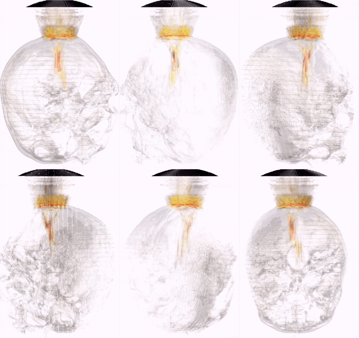
The blood-brain barrier (BBB) restricts the diffusion of microscopic objects, e.g., protecting the brain from infections. However, it also prevents the passage of most therapeutic drugs. BBB disruption can be achieved by using transcranial focused ultrasound and microbubble injection in an effective, non-invasive, transient, localized and safe manner. Thus, the BBB opening enables drug delivering to specific areas of the brain, which is mandatory in research of treatments for neurological disorders as Alzheimer’s or Parkinson’s diseases. At the UMIL we develop simulations and experiments to define a protocol for the transcranial targeting of the hippocampus of an adult human by using a single-element focused transducer. In this line I'm working at the Ultrasound Medical and Industrial Laboratory (UMIL) Universitat Politècnica de València (Spain) and Columbia University (USA) to study the acoustic aberrations produced in transcranial propagation and methods to correct them. We develop simulations and experiments to define a protocol for the transcranial targeting at human hippocampus by using a single-element focused transducers.
In the right animation you can see an example of the transcranial beam propagation generated by a mono-element 500 kHz transducer, obtained by numerical simulation by PhD student Sergio Jiménez-Gambín who is working in this topic (now postdoc at Columbia University). The simulations, based on the k-space method, allow to evaluate the beam aberrations and predict precisely the central nervous system area to be treated.
N. Jiménez, F. Marquet, F. Camarena, E. E. Konofagou, J. Redondo, B. Roig, R. Picó,
Revista de Acustica, 43 (1), pp 5-11, (2012)
P. C. Iglesias, N. Jiménez, E. Konofagou, F. Camarena, J. Redondo
Physics Procedia., 63, pp 103-107, (2014)