Research focus
I work on photoacoustic imaging, magnetomotive ultrasound and ultrasound elastography. These techniques combine acoustic measurements with additional physical mechanisms to explore molecular contrast and the elastic and viscoelastic properties of biological tissues.
Ultrasound has important biomedical applications in both, therapy and imaging. We develop several hybrid ultrasound imaging techniques for biomedical applications combining ultrasound with additional physical mechanisms to go a step beyond standard US imaging techniques. These include photoacoustic imaging, combining laser with ultrasound; and magneto-motive ultrasound, combining magnetic fields, magnetic nanoparticles and ultrasound, to obtain images with molecular contrast. In addition, we are developing ultrasound elastography modalities, combining external forces, torques, and shear deformations with ultrafast ultrasound imaging to map the stiffness of tissues. Finally, we also develop ultrasonic imaging technology to be applied to the field of odontology, quantifying parameters such as the demineralization or to monitor dental implants.
Lines:
Photoacoustic imaging techniques
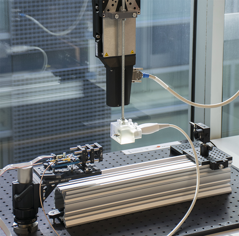
When an ultrashort and intense laser illuminates light-absorbing materials, a part of the absorbed energy produces locally ultrasonic pulses due to thermoelastic effect. Light absorbers become acoustic sources, emmiting ultrasound pulses. These signals can be detected by an ultrasound imaging system can be used to reconstruct an image that take into account the absorbance of the tissue. As the absorbance spectrum varies between tissues and chemical composition it enables the ultrasonic imaging at molecular level.
We have quantified the imaging performance of a large field-of-view optical-resolution photoacoustic-microscopy system using an phased-array detector. The system combines a low-cost pulsed-laser diode with a 128-element linear ultrasound probe. The imaging capabilities of the system are demonstrated generating high-resolution images of tissue-mimicking phantoms containing sub-millimetre ink tubes and an ex vivo rabbit's ear. The results show that dynamic focus DAS beamforming increases and homogenizes SNR along large images, reaching values up to 15 dB compared to an unfocused detector and up to 30 dB compared to out-of-focus regions of the fixed focus configuration. Moreover, the obtained values of gCNR using the DAS beamformer indicate an excellent target visibility, both on phantoms and ex vivo. This strategy makes it possible to scan larger surfaces compared to standard configurations using single-element detectors, paving the way for advanced array-based PAM systems.
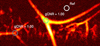
Other ongoing developments include a photoacoustic tomographic system based on a Optical Parametric Oscillator laser tuned at 532 nm and biocompatible nanoparticles acting as a targeted contrast agents.
Related publications
- 2025 · Journal article
Nanoparticles of non-porphyrinic covalent organic frameworks as contrast agents for photoacoustic imaging
Scientific Reports, 15, 33909, 2025
- 2023 · Journal article
Spatial resolution and reconstructed size accuracy using advanced beamformers in linear array-based PAT systems
Photoacoustics, 34, 100576, 2023
- 2023 · Journal article
Imagen fotoacústica empleando nanopartículas de redes orgánicas covalentes (nCOFs) como agente de contraste
Revista de Acústica, 54 (3-4), 28-33, 2023
- 2022 · Journal article
Evaluación del rendimiento de algoritmos de reconstrucción de imágenes para tomografía fotoacústica
Revista de Acústica, 53 (1-2), 17-25, 2022
- 2020 · Journal article
Beamforming for large-area scan and improved SNR in array-based photoacoustic microscopy
Ultrasonics, 111, 106317, 2020
Magnetomotive ultrasound imaging
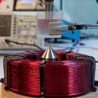
Magneto-Motive ultrasound imaging detects the presence of superparamagnetic nanoparticles through their mechanical responses to an external transient magnetic excitation. Due to their weak diamagnetic properties, normal tissue constituents do not respond to the magnetic field. However, when tissue is labeled with magnetic nanoparticles, it tends to move towards areas of lower magnetic potential. The magnetically-induced displacement within nanoparticles and the tissue associated with them is detectable by ultrasound imaging.
At UMIL we are already developing the first prototype for magnetomotive ultrasound imaging. We started with large particles, which offer the possibility to extract the elasticity of the surrounding medium with great accuracy. The proposed approach is based on the application of a magnetic force to a millimeter-sized steel sphere embedded in the medium and the subsequent monitoring of its dynamical response. A coil is used to create a magnetic field inducing the displacement of the sphere located inside a gelatin phantom. Then, a phased-array system using a 3 MHz ultrasound probe operating in pulse-echo mode is used to track the displacement of the sphere. Experiments were conducted on several samples and repeated as a function of phantom temperature. The dynamical response of the sphere measured experimentally is in good agreement with Kelvin-Voigt theory.
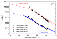
Since the magnetic force is not affected by weak diamagnetic media, our proposal results in an accurate estimation of the force acting on the inclusion. Consequently, the estimated viscoelastic parameters show excellent robustness, and the elastic modulus agrees with the measurements using a quasi-static indentation method, obtaining errors below 10% in the whole temperature range. The use of macroscopic inclusion limits the direct application of this method in a biomedical context, but it provides a robust estimation of the elastic modulus that can be used for material characterization in industrial applications.
Related publications
- 2021 · Journal article
Characterization of viscoelastic media combining ultrasound and magnetic-force induced vibrations on an embedded soft magnetic sphere
IEEE Transactions on Ultrasonics, Ferroelectrics, and Frequency Control, 68 (12), 3540 - 3548, 2021
Elastography
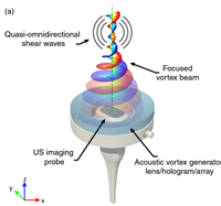
Ultrasonic elastography is a well-established diagnostic tool for pathologies such as liver fibrosis. At the UMIL we are developing novel elastographic techniques using acoustic radiation force and hybrid techniques.
Recently, we presented a novel method of elastography based on acoustic vortices to transfer angular momentum to tissue in addition to linear momentum. Focused vortex beams can push and twist the tissue. In addition, the rotation direction of the applied torque can be dynamically controlled by the modulation of the topological charge of the vortex. The technique results in a robust excitation of shear waves with quasi-omnidirectional radiation pattern and arbitrary waveform, which may have a great impact in imaging performance for elastography.
Related publications
- 2023 · Journal article
Midiendo la elasticidad de tejidos biológicos empleando ultrasonidos
Revista de Acústica, 54 (3-4), 12-19, 2023
- 2020 · Conference contribution
A new elastographic technique using acoustic vortices
IUS 2020: IEEE International Ultrasonics Symposium. Las Vegas, USA. 7-11 September, Oral, 2020
Related conference contributions (1)
- 2024 · Conference contribution
Prostactum: un dispositivo para el examen rectal de la Próstata como método objetivo de detección del Cáncer de Próstata
LXXXVII Congreso Nacional de Urología. Palma de Mallorca, Spain. 12 - 14 Juny 2024, Poster, 2024
Ultrasonic technology applied to odontology
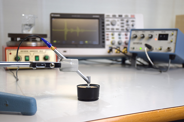
In collaboration with the Valencian Institute of Dental Research (IVIO), within the framework of IVIO-UPVChair, we develop new ultrasonic techniques for monitoring, diagnosis and treatment in the field of dentistry.
We develop ultrasonic characterization methods for guided bone regeneration during implantation in odontology. We aim to characterize the complete regeneration process using ultrasound to guide the treatment, optimize implantation and avoid or detect complications. The use of ultrasound is desirable as it is low cost and non-ionizing radiation, thus, it can be employed to monitor long healing treatments without the risks associated to X-Ray Computed Tomography imaging techniques.
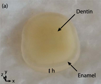
Moreover, ongoing research is developed at UMIL to quantitatively characterize the mechanical properties of teeth, e.g.,
Related publications
- 2021 · Journal article
Ultrasonic monitoring of dentin demineralization
IEEE Transactions on Ultrasonics, Ferroelectrics, and Frequency Control, 68 (3), 570-578, 2021
- 2019 · Journal article
Monitoring the setting of calcium sulphate bone-graft substitute using ultrasonic backscattering
IEEE Transactions on Ultrasonics Ferroelectrics and Frequency Control, 66 (10), 1658-1666, 2019
More publications in this research area
- 2025 · Journal article
Evaluation of a PET Insert for Trimodal Imaging: A Step Towards PET/MRI-Guided Focused Ultrasound
IEEE Transactions on Radiation and Plasma Medical Sciences, early_acccess, 2025
- 2025 · Journal article
Real-time ultrasound shear wave elastography using a local phase gradient
Computer Methods and Programs in Biomedicine, 260, 108529, 2025
- 2015 · Journal article
Macroscopic acousto-mechanical analogy of a microbubble
The Journal of the Acoustical Society of America, 138 (6), 3600-3606, 2015
- 2013 · Journal article
Modeling acoustically driven microbubbles by macroscopic discrete-mechanical analogues
Modelling in Science Education and Learning, 6 (7), 75-87, 2013
Related conference contributions (8)
- 2016 · Conference contribution
Localized nonlinear modes in microbubbles under the action of ultrasound
IUS 2016: IEEE International Ultrasonics Symposium. Tours, France. 18-21 September, Oral, 2016
- 2014 · Conference contribution
Lattice of nonlinear coupled oscillators: An acousto-mechanical analogy of gas microbubble
IUS 2014: IEEE International Ultrasonics Symposium. Chicago, USA. 3 - 6 September, Oral, 2014
- 2014 · Conference contribution
Analogie macroscopique et acousto-mécanique d’une microbulle d’agent de contraste
CFA 2014: 12ème Congrès Français d'Acoustique. Potiers, France. 22 - 25 April, Oral, 2014
- 2013 · Conference contribution
Vibration modes in a pendulums ring: analogy with gas microbubbles surface modes
IUS 2013: IEEE International Ultrasonics Symposium. Prague, Czech Republic. 21-25 July, Oral, 2013
- 2012 · Conference contribution
Spatio-temporal dynamics in a ring of coupled pendula: analogy with bubbles
LENCOS'12: Localized Excitations in Nonlinear Complex Systems, Seville, Spain. 9 - 12 July, Oral, 2012
- 2012 · Conference contribution
Modeling acoustically driven microbubbles by macroscopic discrete-mechanical analogues
III Jornadas de Modelización Matemática. Gandia, Spain. 7 - 8 June, Oral, 2012
- 2011 · Conference contribution
Modulational instability and localized modes for ultrasound contrast microbubbles surface oscillations
IUS 2011: IEEE International Ultrasonics Symposium. Orlando, Florida, USA. 18-21 October, Oral, 2011
- 2011 · Conference contribution
Modulational instability of microbubbles surface modes
ICU 2011: International Congress on Ultrasonics. Gdansk, Poland. 5 - 8 September, Oral, 2011